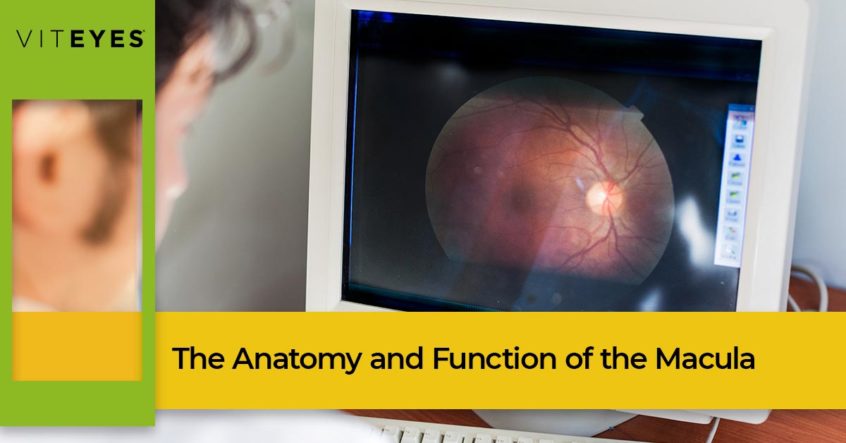
The human eye is such an amazing organ, capable of differentiating approximately 10 million different colors, depth, and shape to offer one of the greatest gifts — sight. Our eyes are considered to be one of our vital organs and sight is considered to be essential to survival. These small organs are only about an inch across and weigh about 0.25 ounce, but incredulously is made up of more than 2 million working parts! In today’s post, we are going to narrow our focus on one of the parts of your eye that can cause significant changes in vision, the macula.
Macular degeneration is a hot topic these days as more awareness has been given to the disease. Macular degeneration is a progressive degenerative eye disease that can cause vision changes and blindness. But, how does this happen, you may ask? Before we can really understand macular degeneration, let’s take a deeper dive into the anatomy and function of the macula.
Anatomy of the Macula
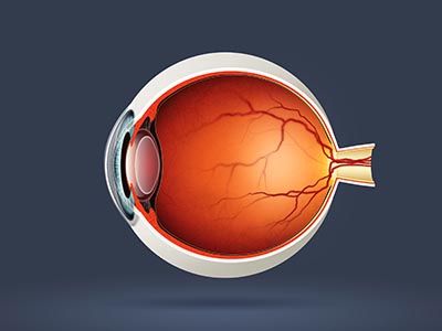
The macula is a very small, but important area located in the center of the retina at the back of your eye. The macula is an oval-shaped pigmented area located near the center of the retina which is only about 5.5 mm. This tiny structure is divided into the umbo, foveola, foveal avascular zone (FAZ), fovea, parafovea, and perifovea areas, each nestled inside the other. Each of these areas plays an important role. The foveola is the concentric ring surrounding the umbo and contains cone cells — photoreceptor nerve cells — that are longer, slimmer, and more densely packed than anywhere else in the eye. The foveal avascular zone is the center of the macula and does not contain retinal blood vessels. The fovea includes the parafovea and perifovea regions and contains closely packed cones.
The Function of the Macula
The macula is responsible for all of our central fine-detailed straight-ahead vision and identifies most of the colors we can see. The macula is what provides clarity and the 20/20 vision we use to focus and identify objects and people. The fovea is thought of to be the most important part of the eye and is responsible for the sharp central vision that allows you to see details and helps you read and drive and is known to be the area of the eye that offers the best visual acuity.

Complications of the Macula
Because of the small size of the macula, it takes very little disturbance to impact your vision. In fact, although macular degeneration gets the most attention, there are many diseases that affect the macula and your vision, including:
- Branch retinal vein occlusion
- Central retinal vein occlusion
- Central serous retinopathy
- Choroidal neovascular membranes
- Cytomegalovirus retinitis
- Diabetic retinopathy
- Histoplasmosis
- Macular degeneration
- Macular edema
- Macular hole
- Macular pucker
- Macular telangiectasia
- Retinal detachment
- Retinitis pigmentosa
- Retinoblastoma
- Retinopathy of prematurity
- Stargardt disease
- Usher syndrome
As you can gather from this list, many diseases of the macula originate in the retina, where the macula rests. A good majority of these diseases, including macular degeneration, are caused by abnormal blood flow or damage to the blood vessels of the retina.
Although it is a tiny structure, the macula of your eyes provides the sight you cherish. It is important to protect your macula, retina, and total eye health to support good vision for the duration of your life. That’s where Viteyes comes in. We are an eye vitamin company providing supplements that support optimal macular health. Learn more about macular health in our online resource and shop our entire line of eye supplements online today.